「資料集・リンク」

「専門家向けデータベース」
by MOE and Chemicals Evaluation and Research Institute, Japan (CERI)
- Top page of Atlas of Medaka Gonadal Histology
- About Medaka (Oryzias latipes)
- Normal Development and Gonadal Histology in Medaka
- Atlas of Gonadal Histopathology in Medaka Exposed to Endocrine Disruptors
- Effect of estrogen exposure
- Effect of androgen exposure
- Effect of anti-androgen exposure
Atlas of Gonadal Histopathology in Medaka Exposed to Endocrine Disruptors
Effect of estrogen exposure
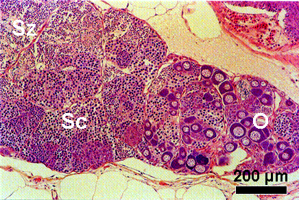
Testis-ova
Testis-ova from a fish exposed to 224 µg/L of 4-tert-pentylphenol from fertilized egg to 61-d posthatch (Bouin, H&E).
O: oocyte, Sc: spermatocyte, Sz: spermatozoa.
Oocytes appear in clusters within the testicular tissue. Numerous spermatocytes and spermatozoa are still present in a compacted mass in this section.

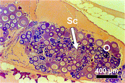
Progressed testis-ova
Testis-ova from a fish exposed to 931 µg/L of 4-tert-pentylphenol from fertilized egg to 61-d posthatch (Bouin, H&E).
O: oocyte, Sc: spermatocyte, Sz: spermatozoa.
A more progressed testis-ova. Almost the entire area is composed of oocytes, accompanying small testicular tissues interspersed with a few spermatocytes.

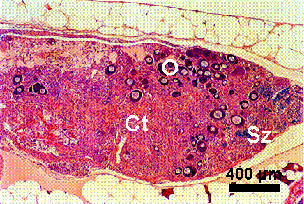
Testis-ova & connective tissue
Testis-ova from a fish exposed to 931 µg/L of 4-tert-pentylphenol from fertilized egg to 61-d posthatch (Bouin, H&E).
O: oocyte, Ct: connective tissues, Sz: spermatozoa.
Connective tissues develop well in a testis-ova specimen with a few spermatozoa.

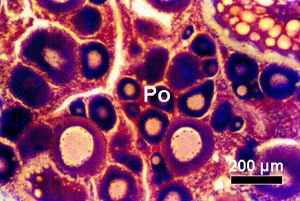
Regressed ovary
Ovary from an adult female fish exposed to 488 ng/L of ethinylestradiol for 3 weeks (Bouin, H&E,).
Po: previtellogenic oocyte
Many previtellogenic oocytes exist in this specimen, suggesting regressed ovary.


