X-rays for Medical Use and Generators
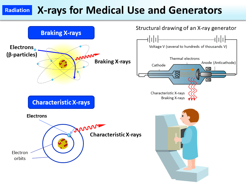
X-ray examination uses X-rays generated in X-ray tubes. A high voltage is applied between a cathode and an anode (tungsten, molybdenum, copper, etc.) inside an X-ray tube so that thermal electrons migrate from the cathode to the anode in a vacuum at high speed. X-rays generated when the direction of propagation of the thermal electrons changes as they are attracted to the nucleus of the anode are called braking X-rays. When an electron is ejected from the inner electron orbit of the anode nucleus, another electron migrates (transitions) to this vacancy from the outer electron orbit. X-rays generated thereby are called characteristic X-rays. Most of the X-rays generated in X-ray tubes are braking X-rays.
Generation of X-rays stops when the X-ray tube is switched off.
X-ray generators used in the field of medicine are either for diagnosis or for treatment. The energy and amount of X-rays are adjusted to match the purpose of imaging and the part to be imaged. In chest roentgenography (diagnosis), the amount of radiation a patient receives in one imaging session is approx. 0.06 mSv.
- Included in this reference material on March 31, 2016