Thyroid Ultrasound Examination: Outline (3/3)
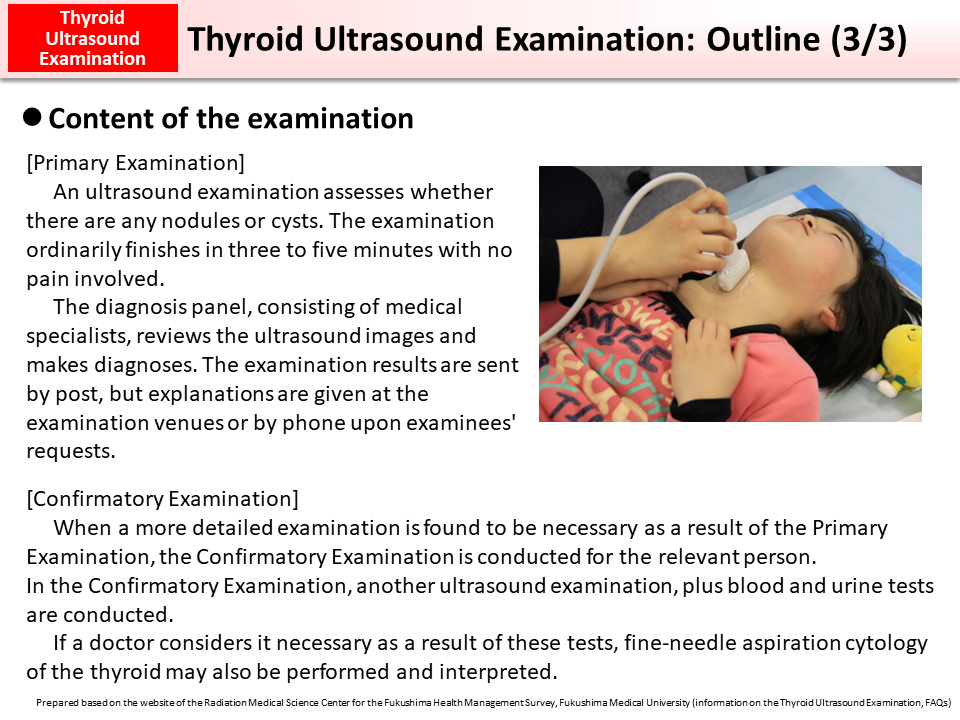
An ultrasound examination is conducted with an examinee lying on his/her back. A doctor places an ultrasonic probe with jelly on its tip over the examinee's thyroid (located around the base of the neck) and examines whether there are any cysts or nodules while moving the probe over the examinee's skin.
The examination ordinarily finishes in three to five minutes with no pain involved.
Definitive diagnoses from the Primary Examination are not made at the venues. In order to make comprehensive and objective judgments, ultrasound images are later reviewed by a panel of medical specialists. This is to ensure a consistently high level of diagnostic accuracy throughout the Fukushima Health Management Survey.
The sizes of nodules and cysts mentioned above are reference values for making diagnoses. If any nodules or cysts found in ultrasound images are suspected to be malignant, the case is designated as Grade B irrespective of the sizes of the nodules or cysts and the Confirmatory Examination is recommended.
In the Confirmatory Examination, a more accurate ultrasound examination, plus blood and urine tests, are conducted. If, as a result of these tests, a doctor considers it necessary, fine-needle aspiration cytology, an examination of a sample tissue taken from the person's thyroid, may also be conducted.
- Included in this reference material on March 31, 2016
- Updated on March 31, 2019