X-rays for Medical Use and Generators
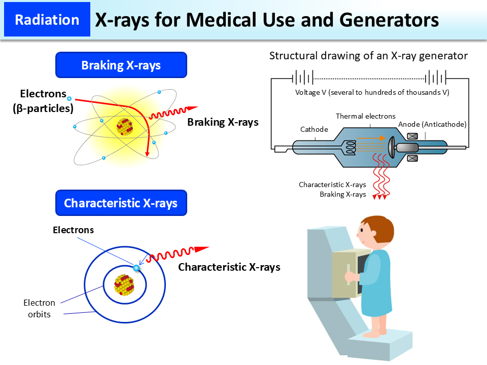
X-ray examination uses X-rays generated in X-ray tubes. A high voltage is applied between a cathode and an anode (tungsten, molybdenum, copper, etc.) inside an X-ray tube so that thermal electrons migrate from the cathode to the anode in a vacuum at high speed. X-rays generated when the direction of propagation of the thermal electrons changes as they are attracted to the nucleus of the anode are called braking X-rays. When an electron is ejected from the inner electron orbit of the anode nucleus, another electron migrates (transitions) to this vacancy from the outer electron orbit. X-rays generated thereby are called characteristic X-rays. Most of the X-rays generated in X-ray tubes are braking X-rays.
Generation of X-rays stops when the X-ray tube is switched off.
X-ray generators used in the field of medicine are either for diagnosis or for treatment. The energy and amount of X-rays are adjusted to match the purpose of imaging and the part to be imaged. In chest roentgenography (diagnosis), the amount of radiation a patient receives in one imaging session is approx. 0.06 mSv.
(Related to p.63 of Vol. 1, “Exposure Dose from Natural and Artificial Radiation,” and p.76 of Vol. 1, “Radiation Doses from Medical Diagnosis”)
- Included in this reference material on March 31, 2016
- Updated on March 31, 2022